


Explore the Practice Resource Centre

EXPLORE THE PRACTICE RESOURCE CENTRE – we are pleased to share the new PRC promotional video. In just over two minutes, we highlight the benefits of the site for seasoned practitioners and those just starting their career, how to easily submit ideas and resources, how to create a custom reading list, and much more.
Glaucoma OCT Interpretation: 101
Glaucoma OCT Interpretation is an online, Section 3 accredited course available at EyeCarePD.com. A game-based approach is applied to learning OCT interpretation. This course covers commonly encountered OCT presentations of glaucoma as seen on standard optic nerve head and retinal nerve fiber layer scan protocols with the purpose of improving interpretation skills using perceptual learning strategies.
Getting started is easy:
- Using the Google Chrome browser, navigate to https://eyecarepd.com/catalog/glaucoma-group/
- Select Glaucoma OCT Interpretation 101 (COS Accredited) and add the product to the cart
- At checkout, use the coupon code ECPDCOSGLAUCOMA to set the price to 0.
- Your course is now available under My Courses at the top of the screen.
Learning Objectives:
- Identify segmentation errors and their role in OCT scan interpretation
- Utilize varying sections of the OCT report including tabular data, sector analysis and graphical displays
- Interact with expert interpretation in order to compare their findings in selected cases
Accreditation
This activity is an Accredited Self-Assessment Program (Section 3) as defined by the Maintenance of Certification program of the Royal College of Physicians and Surgeons of Canada. This web-based lesson was approved by the COS on October 23rd, 2020 and expires October 2023. Remember to visit MAINPORT to record your learning and outcomes. You may claim a maximum of 1 hour (credits are automatically calculated).
This activity was co-developed with EyeCarePD and the Canadian Ophthalmological Society (COS) and was planned to achieve scientific integrity, objectivity and balance.
Access Details
To participate in this course visit https://eyecarepd.com/catalog/glaucoma-group/

Advanced Therapeutic Options for Ocular Surface Disease: Blood-Derived Products, Insulin Eye Drops, and Human Amniotic Membrane
COS Practice Resource Centre General Information for Patients and Providers
Ergonomics and Injury Prevention Webinar
Save the Date!
Date: Thursday, October 8, 2026, 7:00 pm ET
Title: Keeping You in the Loupe: A Closer Look at Ergonomics in the Operating Room
Intravenous Acetazolamide Shortage
The Atlantic Eye Symposium
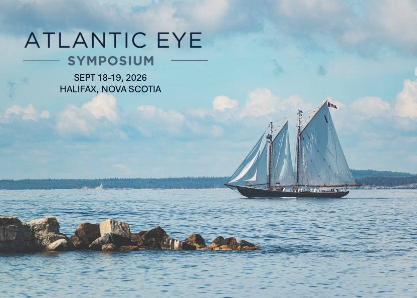
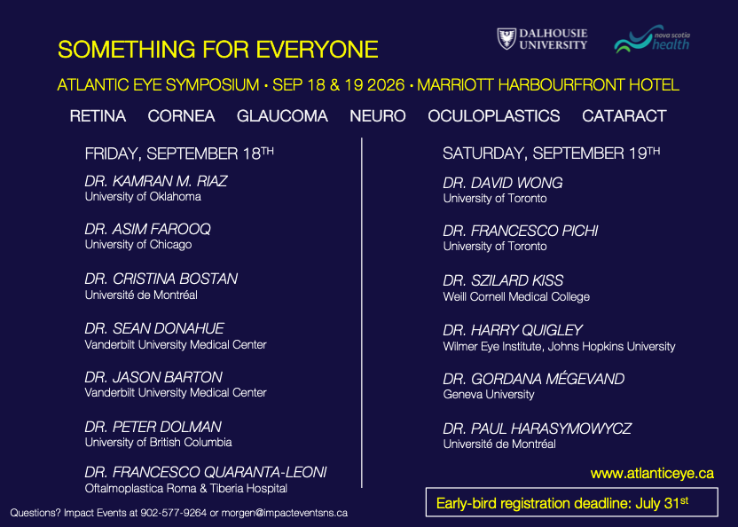
The Atlantic Eye Symposium is an accredited educational conference geared to address the needs of the comprehensive ophthalmologist. There will be 13 visiting speakers covering a variety of topics including cataract, cornea, retina, glaucoma, neuro-ophthalmology, and oculoplastics.
This two-day meeting will be held on the Halifax waterfront at the Harbourfront Marriott Hotel on September 18th and 19th, 2026. The informal setting will facilitate dialogue and interaction with the speakers and your colleagues. You can expect innovative talks and exhibitions addressing current issues in the field of ophthalmology.
EyeNovation Webinar: INTRA-KER: A Novel Concept for a Hybrid Keratoprosthesis

Did you miss the webinar? Not to worry, a recording is available for you below!
Our EyeNovation webinar featured Dr. Massimo Busin, who presented on INTRA-KER: A Novel Concept for a Hybrid Keratoprosthesis.
This session explored the concept of INTRA-KER, a hybrid keratoprosthesis that integrates a synthetic optical device with a biologic scaffold, offering practical insights and clinical perspectives relevant to ophthalmologists and trainees.

Massimo Busin, MD, is the chairman of the Department of Ophthalmology at “Ospedali Privati Forlì” in Forlì (Italy) and is also a Professor of Ophthalmology at the University of Bonn (Germany), as well as and Adjunct Associate Clinical Professor at LSU (New Orleans, USA). Since 2017 he has been appointed Professor of Ophthalmology and Director of the Residency program of the Department of Ophthalmology in Ferrara (Italy)
Dr. Busin has received from the AAO (American Academy of Ophthalmology) the “Honor Award” in 1993, the “Senior Honor Achievemt” award in 2003, the “Life Achievement” award in 2013 and the “Special Recognition Award” in 2015; from SOI (Società Oftalmologica Italiana) the Gold Medal “Maestro dell’Oftalmologia” in 2012. He has delivered several eponimal lectures and has also received several prizes for both videos and lectures delivered at international meetings, among which the AAO, the ESCRS, the DOC, the ASCRS, the SOI. He has authored more than 250 peer-reviewed papers, and 24 book chapters.
Learning Objectives
By the end of this session, participants will be able to:
- Understand the foundational concept, key surgical principles as well as clinical indications of INTRA-KER
CanMEDS Role: Medical Expert
Representation Circle: Surgical Learning, Research and Mentorship – Recording
Did you miss the webinar? Not to worry, a recording is available for you below!
Our inaugural Representation Circle webinar featured Dr. Prithvi Mruthyunjaya, who presented on Surgical Learning, Research and Mentorship.
This interactive session explored mentorship, surgical learning, and research development in ophthalmology training, while creating space for meaningful discussion and shared experiences across the ophthalmology community. The session also included a live Q&A with participants.

Dr. Prithvi Mruthyunjaya is the Alan Adler Professor of Ophthalmology, member of the Vitreoretinal Surgery Service, and Director of Ocular Oncology at the Byers Eye Institute at Stanford. As Director of the Vitreoretinal Surgery and Medical Retina Fellowships, he has helped build a highly regarded academic-focused retina training program and has trained more than 50 retina and ocular oncology fellows over the past 20 years. He has coauthored over 230 peer-reviewed publications and is internationally recognized for his contributions to surgical education, mentorship, and vitreoretinal training.
Dr. Awwad serves as Associate Editor of the Journal of Refractive Surgery and sits on the Editorial Board of the American Journal of Ophthalmology. His work has earned numerous international distinctions, including the George Waring Medal for Editorial Excellence and the AAO Achievement Award. He has also contributed innovative analytical tools and formulas to major ophthalmic platforms and holds patents in artificial intelligence and computer vision related to ophthalmic surgery.
Learning Objectives
By the end of this session, participants were able to:
- Review elements of organizing surgical learning in residency
- Understand concepts of research development in residency
EyeNovation Webinar: The Evolving Landscape of CAIRS – Recording
Did you miss the webinar? Not to worry, a recording is available for you below!
Our EyeNovation webinar featured Dr. Awwad, who presented on The evolving landscape of CAIRS.
This session explored the latest developments in CAIRS, including evolving surgical techniques, preparation methods, and clinical outcomes, offering valuable, practice-oriented insights for ophthalmologists and trainees.

Dr. Shady Awwad is Professor of Ophthalmology and Head of the Cornea & Refractive Surgery Division at the American University of Beirut Medical Center, where he also founded the Refractive Surgery Fellowship Program. He is internationally recognized for his expertise in refractive and keratoconus surgery and has authored more than 130 peer-reviewed publications.
Dr. Awwad serves as Associate Editor of the Journal of Refractive Surgery and sits on the Editorial Board of the American Journal of Ophthalmology. His work has earned numerous international distinctions, including the George Waring Medal for Editorial Excellence and the AAO Achievement Award. He has also contributed innovative analytical tools and formulas to major ophthalmic platforms and holds patents in artificial intelligence and computer vision related to ophthalmic surgery.
Learning Objectives
By the end of this session, participants will be able to:
- Introduction to CAIRS, their outcomes and their mechanism of action
- Insights into the changing trends in CAIRS preparation, insertion, and surgical techniques.
Keratoprosthesis: Best Practices – Preoperative, Operative and Postoperative
University of Toronto and Wills Eye Hospital are proud to present the next installment of the Cornea Surgical Video Learning Series
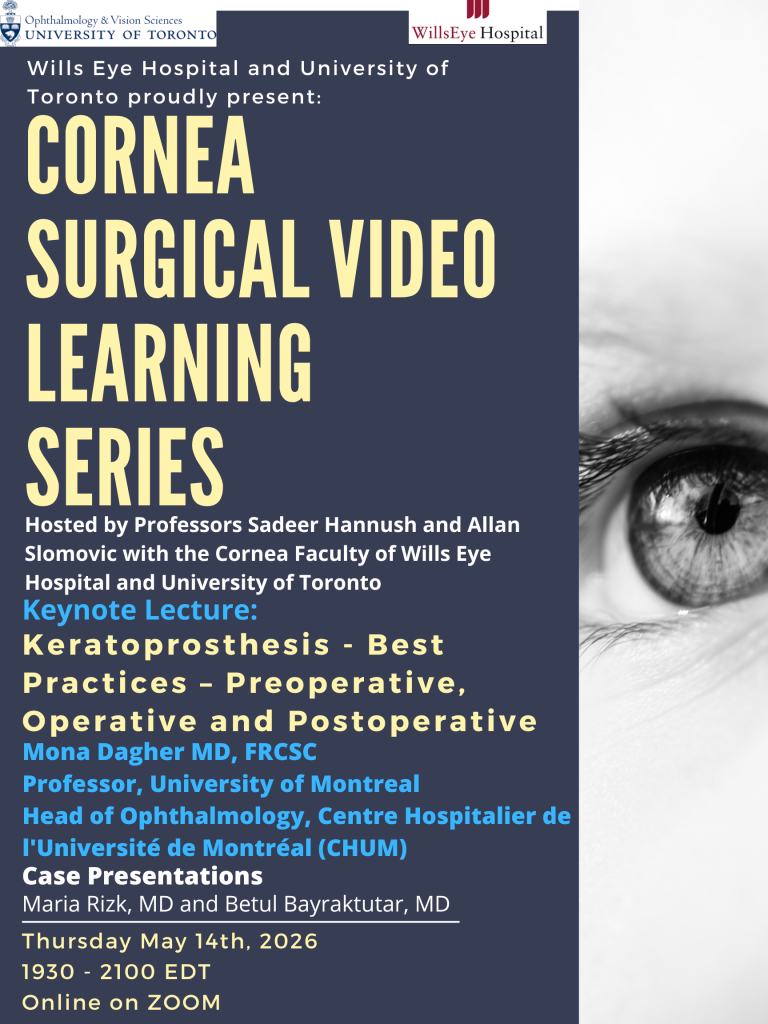
Date: Thursday, May 14th, 2026
Time: 1930 – 2100 PM EDT
Co-moderated by Professors Sadeer Hannush (Wills Eye Hospital) and Allan Slomovic (University of Toronto).
Keynote Lecturer
Mona Dagher MD, FRCSC
Professor, University of Montreal
Head of Ophthalmology, Centre Hospitalier de l’Université de Montréal (CHUM)
Guest Panellists
Neera Singal MD, FRCSC and Clara Chan MD, FRCSC (University of Toronto)
Marisa Schoen MD & Zeba Syed MD (Wills Eye Hospital)
Surgery Case Presentations
Maria Rizk MD, FRCSC (Université de Montréal)
Betul Bayraktutar MD, DO (Wills Eye Hospital)
You are invited to a Zoom webinar. You must register in order to join and receive your CPD.
https://us02web.zoom.us/webinar/register/WN_CjpTHmDnRs-z1kvSbjvEGA#
After registering, you will receive a confirmation email containing information about joining the webinar.
The evening will as usual be recorded and placed online for viewing.
If you would like to view prior sessions in the series, please visit:
https://ophthalmology.utoronto.ca/subspecialty-rounds
CME credits are available for the following:
· Royal College Maintenance of Certification Section 1: 1.5 hours (1.5 Section 1 hours per session)
· American Medical Association Category 1: 1.5 credits (1.5 Category 1 credits per session)
· European Union for Medical Specialists UEMS-EACCME®: 1.5 credits (1.5 ECMEC credits per session)
Ergonomics: Journal Articles & Editorials
- Bolis M, Garg A, Chan B. Real-time postural feedback to optimize ergonomics and musculoskeletal health in ophthalmology residents: a Canadian pilot quality improvement study. Cureus 2025;17(7):e88736. DOI: 10.7759/cureus.88736 https://pubmed.ncbi.nlm.nih.gov/40717881/
- Buchmann W, Fortineau E, Ribaute E, Brezin A. [Musculoskeletal problems in ophthalmology, risk factors and solutions: case studies from two French practices]. Journal Français d’Ophtalmologie 2025;48(7):104578. DOI: 10.1016/j.jfo.2025.104578 https://pubmed.ncbi.nlm.nih.gov/40669315/
- Ismail MM, Fayyadh RA, Hassanein DH, et al. Prevalence and risk factors of musculoskeletal pain among pediatric ophthalmologists and strabismologists in the Middle East: a cross-sectional study. Journal of Pediatric Ophthalmology and Strabismus 2026;63(1):65-73. DOI: 10.3928/01913913-20250701-01 https://pubmed.ncbi.nlm.nih.gov/40736052/
- Kaur H, Xie JS, Lusterio A, et al. A scoping review of ergonomics in ophthalmology: working in pain. American Journal of Ophthalmology 2026;282:433-57. DOI: 10.1016/j.ajo.2025.11.005 https://pubmed.ncbi.nlm.nih.gov/41242593/
- Nishant P, Singh A, Morya AK, et al. Manual small incision cataract surgery: an ergonomic solution to tackle cataract backlog and challenging situations. World Journal of Methodology 2025;15(4):104529. DOI: 10.5662/wjm.v15.i4.104529 https://pubmed.ncbi.nlm.nih.gov/40900862/
- Sather RN, 3rd, Moon JY, Romano F, et al. The ergonomic evaluation of attendings and trainees across the vitreoretinal service as measured by a wearable device. Ophthalmic Surgery, Lasers and Imaging Retina 2025;56(2):80-5. DOI: 10.3928/23258160-20240906-01 https://pubmed.ncbi.nlm.nih.gov/39311565/
- Sinha S, Nishant P, Sinha RK. Experience and challenges in intraocular surgery in patients with kyphosis: a cross-sectional survey among practicing ophthalmologists. Cureus 2025;17(8):e89490. DOI: 10.7759/cureus.89490 https://pubmed.ncbi.nlm.nih.gov/40918920/
- Tanya SM, Kulbay M, Kherani A, et al. Ergonomic impact of electronic medical records on Canadian eye care providers: results from the Canadian ophthalmic practitioners ergonomics survey. Canadian Journal of Ophthalmology 2026. DOI: 10.1016/j.jcjo.2026.01.008 https://pubmed.ncbi.nlm.nih.gov/41620202/
- Xie JS, Kaur H, Lusterio A, et al. A scoping review of ergonomics in ophthalmology: working smarter. American Journal of Ophthalmology 2026;281:249-69. DOI: 10.1016/j.ajo.2025.08.053 https://pubmed.ncbi.nlm.nih.gov/40889624/
- Aloqab A, Alturkistany W, Ali HMN. The relationship between surgical loupes usage, workplace ergonomics, and musculoskeletal disorders among Saudi ophthalmologists. Saudi Journal of Ophthalmology. 2024; 3;39(2):166-173. DOI: 10.4103/sjopt.sjopt_276_23. https://pubmed.ncbi.nlm.nih.gov/40642356/
- Morrison AK, Kumar S, Amin A, Urban M, Kleinman B. An ergonomic risk assessment of ophthalmology residents using the Rapid Entire Body Assessment (REBA) scale. Cureus 2024;16(2):e53698. DOI: 10.7759/cureus.53698. https://pubmed.ncbi.nlm.nih.gov/38455825/
- Kamei M, Suzuki H, Terayama H, et al. Ergonomic benefit using heads-up display compared to conventional surgical microscope in Japanese ophthalmologists. PLoS One 2024;19(5):e0297461. DOI: 10.1371/journal.pone.0297461. https://pubmed.ncbi.nlm.nih.gov/38776346/
- Barrios EL, Polcz VE, Hensley SE, et al. A narrative review of ergonomic problems, principles, and potential solutions in surgical operations. Surgery 2023;174(2):214-221. DOI: 10.1016/j.surg.2023.04.003. https://pubmed.ncbi.nlm.nih.gov/37202309/
- Fouzdar Jain S, Akhter S, Ishihara R, Siddicky S, High R, Suh DW. The prevalence of work-related musculoskeletal disease among pediatric ophthalmologists. Clinical Ophthalmology 2022;16:833-840. DOI: 10.2147/OPTH.S343155. https://pubmed.ncbi.nlm.nih.gov/35330751/
- Cerier E, Hu A, Goldring A, Rho M, Kulkarni SA. Ergonomics Workshop Improves Musculoskeletal Symptoms in General Surgery Residents. Journal of Surgical Research 2022;280:567-574. DOI: 10.1016/j.jss.2022.06.014.https://pubmed.ncbi.nlm.nih.gov/35787315/
- Albanesi B, Piredda M, Bravi M, et al. Interventions to prevent and reduce work-related musculoskeletal injuries and pain among healthcare professionals. A comprehensive systematic review of the literature. Journal of Safety Research 2022;82:124-143. DOI: 10.1016/j.jsr.2022.05.004. https://pubmed.ncbi.nlm.nih.gov/36031239/
- Aaron KA, Vaughan J, Gupta R, et al.The risk of ergonomic injury across surgical specialties. PLoS One 2021;16(2):e0244868. DOI: 10.1371/journal.pone.0244868. https://pubmed.ncbi.nlm.nih.gov/33561117/
- Schechet SA, DeVience E, DeVience S, Shukla S, Kaleem M. Survey of musculoskeletal disorders among US ophthalmologists. Digital Journal of Ophthalmology 2021;26(4):36-45. DOI: 10.5693/djo.01.2020.02.001. https://pubmed.ncbi.nlm.nih.gov/33867881/
- Koshy K, Syed H, Luckiewicz A, Alsoof D, Koshy G, Harry L. Interventions to improve ergonomics in the operating theatre: A systematic review of ergonomics training and intra-operative microbreaks. Annals of Medicine & Surgery (Lond) 2020;55:135-142. DOI: 10.1016/j.amsu.2020.02.008. https://pubmed.ncbi.nlm.nih.gov/32477512/
- Betsch D, Gjerde H, Lewis D, Tresidder R, Gupta RR. Ergonomics in the operating room: it doesn’t hurt to think about it, but it may hurt not to! Canadian Journal of Ophthalmology 2020;55(3 Suppl 1):17-21. DOI: 10.1016/j.jcjo.2020.04.004. https://pubmed.ncbi.nlm.nih.gov/32448408/
- Weng CY, Hariprasad SM, Leiderman YI. Ergonomics in retina. Ophthalmic Surgery, Lasers and Imaging Retina 2019;50(9):537-542. DOI: 10.3928/23258160-20190905-01. https://pubmed.ncbi.nlm.nih.gov/31589750/
- Epstein S, Tran BN, Capone AC, et al. The current state of surgical ergonomics education in U.S. surgical training: a survey study. Annals of Surgery 2019;269(4):778-784. DOI: 10.1097/SLA.0000000000002592. https://pubmed.ncbi.nlm.nih.gov/29381528/
- Diaconita V, Uhlman K, Mao A, Mather R. Survey of occupational musculoskeletal pain and injury in Canadian ophthalmology. Canadian Journal of Ophthalmology 2019;54(3):314-322. DOI: 10.1016/j.jcjo.2018.06.021. https://pubmed.ncbi.nlm.nih.gov/31109470/
- Bonafede L, Kazmierczak L, Siddicky SF, Gunton KB. Ergonomics in strabismus surgery. Current Opinion in Ophthalmology 2019;30(5):331-336. DOI: 10.1097/ICU.0000000000000594. https://pubmed.ncbi.nlm.nih.gov/31313751/
- Ratzlaff TD, Diesbourg TL, McAllister MJ, von Hacht M, Brissette AR, Bona MD. Evaluating the efficacy of an educational ergonomics module for improving slit lamp positioning in ophthalmology residents. Canadian Journal of Ophthalmology 2019;54(2):159-163. DOI: 10.1016/j.jcjo.2018.05.016. https://pubmed.ncbi.nlm.nih.gov/30975337/
- Kaup S, Shivalli S, Kulkarni U, Arunachalam C. Ergonomic practices and musculoskeletal disorders among ophthalmologists in India: An online appraisal. European Journal of Ophthalmology 2020;30(1):196-200. DOI: 10.1177/1120672118815107. https://pubmed.ncbi.nlm.nih.gov/30474398/
- Venkatesh R, Kumar S. Back pain in ophthalmology: National survey of Indian ophthalmologists. Indian Journal of Ophthalmology 2017;65(8):678-682. DOI: 10.4103/ijo.IJO_344_17. https://pubmed.ncbi.nlm.nih.gov/28820152/
- Shaw C, Bourkiza R, Wickham L, McCarthy I, McKechnie C. Mechanical exposure of ophthalmic surgeons: a quantitative ergonomic evaluation of indirect ophthalmoscopy and slit-lamp biomicroscopy. Canadian Journal of Ophthalmology 2017;52(3):302-307. DOI: 10.1016/j.jcjo.2016.09.011. https://pubmed.ncbi.nlm.nih.gov/28576213/
- 2Honavar SG. Head up, heels down, posture perfect: Ergonomics for an ophthalmologist. Indian Journal of Ophthalmology 2017;65(8):647-650. DOI: 10.4103/ijo.IJO_711_17. https://pubmed.ncbi.nlm.nih.gov/28820146/
- Sivak-Callcott JA, Mancinelli CA, Nimbarte AD. Cervical occupational hazards in ophthalmic plastic surgery. Current Opinion in Ophthalmology 2015;26(5):392-8. DOI: 10.1097/ICU.0000000000000182. https://pubmed.ncbi.nlm.nih.gov/26247136/
- Alrashed WA. Ergonomics and work-related musculoskeletal disorders in ophthalmic practice. Imam Journal of Applied Sciences 2016;1(2):48-63. DOI: 10.4103/ijas.ijas_24_16. https://journals.lww.com/ijas/fulltext/2016/01020/ergonomics_and_work_related_musculoskeletal.2.aspx
- Hyer JN, Lee RM, Chowdhury HR, Smith HB, Dhital A, Khandwala M. National survey of back & neck pain amongst consultant ophthalmologists in the United Kingdom. International Ophthalmology 2015;35(6):769-775. DOI: 10.1007/s10792-015-0036-z. https://pubmed.ncbi.nlm.nih.gov/25609503/
- Herzog NV, Beharic RV, Beharic A, Buchmeister B. Ergonomic analysis and simulation in department of ophthalmology. Procedia Manufacturing 2015;3:128-135. DOI: 10.1016/j.promfg.2015.07.117. https://www.researchgate.net/publication/283960343_Ergonomic_Analysis_and_Simulation_in_Department_of_Ophthalmology
- Fethke NB, Schall MC, Determan EM, Kitzmann AS. Neck and shoulder muscle activity among ophthalmologists during routine clinical examinations. International Journal of Industrial Ergonomics 2015;49:53-59. DOI: 10.1016/j.ergon.2015.06.001. https://www.sciencedirect.com/science/article/abs/pii/S0169814115300019
- Hedge A, James T. Detrimental Effects of an electronic health records system on musculoskeletal symptoms among health professionals. Proceedings of the Human Factors and Ergonomics Society Annual Meeting 2014;58(1):773-777. DOI: 10.1177/1541931214581141. https://www.researchgate.net/publication/271722221_Detrimental_Effects_of_an_Electronic_Health_Records_System_on_Musculoskeletal_Symptoms_among_Health_Professionals#:~:text=Exposure%20to%20blood%20and%20body,of%20health%20practitioners%20surveyed%20reported
- Mehta S, Hubbard GB, 3rd. Avoiding neck strain in vitreoretinal surgery: an ergonomic approach to indirect ophthalmoscopy and laser photocoagulation. Retina 2013;33(2):439-41. DOI: 10.1097/IAE.0b013e318276cbca. https://pubmed.ncbi.nlm.nih.gov/23190927/
- Dorion D, Darveau S. Do micropauses prevent surgeon’s fatigue and loss of accuracy associated with prolonged surgery? An experimental prospective study. Annals of Surgery 2013;257(2):256-9. DOI: 10.1097/SLA.0b013e31825efe87. https://pubmed.ncbi.nlm.nih.gov/22824853/
- Marx JL. Ergonomics: back to the future. Ophthalmology 2012;119(2):211-2. DOI: 10.1016/j.ophtha.2011.09.001. https://pubmed.ncbi.nlm.nih.gov/22305308/
- Kitzmann AS, Fethke NB, Baratz KH, Zimmerman MB, Hackbarth DJ, Gehrs KM. A survey study of musculoskeletal disorders among eye care physicians compared with family medicine physicians. Ophthalmology 2012;119(2):213-20. DOI: 10.1016/j.ophtha.2011.06.034. https://pubmed.ncbi.nlm.nih.gov/21925736/
- Theou O, Soon Z, Filek S, et al. Changing the sheets: a new system to reduce strain during patient repositioning. Nursing Research 2011;60(5):302-8. DOI: 10.1097/NNR.0b013e318225b8aa. https://pubmed.ncbi.nlm.nih.gov/21873921/
- Sivak-Callcott JA, Diaz SR, Ducatman AM, Rosen CL, Nimbarte AD, Sedgeman JA. A survey study of occupational pain and injury in ophthalmic plastic surgeons. Ophthalmic Plastic & Reconstructive Surgery 2011;27(1):28-32. DOI: 10.1097/IOP.0b013e3181e99cc8. https://pubmed.ncbi.nlm.nih.gov/20859236/
- Dhimitri KC, McGwin G Jr., McNeal SF, et al. Symptoms of musculoskeletal disorders in ophthalmologists. American Journal of Ophthalmology 2005;139(1):179-81. DOI: 10.1016/j.ajo.2004.06.091. https://pubmed.ncbi.nlm.nih.gov/15652844/
- Piccoli B, Committee IS. A critical appraisal of current knowledge and future directions of ergophthalmology: consensus document of the ICOH Committee on ‘Work and Vision’. Ergonomics 2003;46(4):384-406. DOI: 10.1080/0014013031000067473. https://pubmed.ncbi.nlm.nih.gov/12637175/
- Wallace RB, 3rd. The 45 degree tilt: improvement in surgical ergonomics. Journal of Cataract & Refractive Surgery 1999;25(2):174-6. DOI: 10.1016/s0886-3350(99)80122-9. https://pubmed.ncbi.nlm.nih.gov/9951660/
- Chatterjee A, Ryan WG, Rosen ES. Back pain in ophthalmologists. Eye (Lond) 1994;8 ( Pt 4):473-4. DOI: 10.1038/eye.1994.112. https://pubmed.ncbi.nlm.nih.gov/7821477/