


Explore the Practice Resource Centre
EXPLORE THE PRACTICE RESOURCE CENTRE – we are pleased to share the new PRC promotional video. In just over two minutes, we highlight the benefits of the site for seasoned practitioners and those just starting their career, how to easily submit ideas and resources, how to create a custom reading list, and much more.
Glaucoma OCT Interpretation: 101
Glaucoma OCT Interpretation is an online, Section 3 accredited course available at EyeCarePD.com. A game-based approach is applied to learning OCT interpretation. This course covers commonly encountered OCT presentations of glaucoma as seen on standard optic nerve head and retinal nerve fiber layer scan protocols with the purpose of improving interpretation skills using perceptual learning strategies.
Getting started is easy:
- Using the Google Chrome browser, navigate to https://eyecarepd.com/catalog/glaucoma-group/
- Select Glaucoma OCT Interpretation 101 (COS Accredited) and add the product to the cart
- At checkout, use the coupon code ECPDCOSGLAUCOMA to set the price to 0.
- Your course is now available under My Courses at the top of the screen.
Learning Objectives:
- Identify segmentation errors and their role in OCT scan interpretation
- Utilize varying sections of the OCT report including tabular data, sector analysis and graphical displays
- Interact with expert interpretation in order to compare their findings in selected cases
Accreditation
This activity is an Accredited Self-Assessment Program (Section 3) as defined by the Maintenance of Certification program of the Royal College of Physicians and Surgeons of Canada. This web-based lesson was approved by the COS on October 23rd, 2020 and expires October 2023. Remember to visit MAINPORT to record your learning and outcomes. You may claim a maximum of 1 hour (credits are automatically calculated).
This activity was co-developed with EyeCarePD and the Canadian Ophthalmological Society (COS) and was planned to achieve scientific integrity, objectivity and balance.
Access Details
To participate in this course visit https://eyecarepd.com/catalog/glaucoma-group/

Protected: Fuchs’ Module Testing Page
Module Disclosure Page
Scientific Planning Committee
Dr. Parnian Arjmand – Chair
I have a relationship with a for-profit and/or non-profit organization to diclose:
- Any direct financial payments, including receipt of honoraria: COS
- Membership on advisory boards or speakers’ bureaus: Roche, Bayer, Apobiologix
Dr. Vlad Diaconita – Member
I have a relationship with a for-profit and/or non-profit organization to disclose:
- Any direct financial payments, including receipt of honoraria: Voiant LLC Consultant
- Membership on advisory boards or speakers’ bureaus: Biogen, Roche
Dr. Bryon McKay – Member
I do not have a relationship with a for-profit and/or non-profit organization to disclose.
Dr. Miral Mehta-Shounak – Member
I do not have a relationship with a for-profit and/or non-profit organization to disclose.
DOVS CORNEA ROUNDS – Pentacam Pearls for Refractive Surgery Screening – VIA ZOOM
Date: Thursday June 26, 2025
Time: 5:00 PM, ET
Title: Pentacam Pearls for Refractive Surgery Screening
Speaker: Dr. Nir Sorkin
Zoom Link to Register: CLICK HERE
After registering, you will receive a confirmation email containing information about joining the webinar.
The VPP Grand Rounds Local and Visiting Professor Program is a self-approved group learning activity (Section 1) as defined by the Maintenance of Certification program of The Royal College of Physicians and Surgeons of Canada. You will be able to claim 1 credit for this round.
2025 COS Annual Meeting ePosters
The 2025 COS Annual Meeting was held from June 19th to 22nd, 2025 in the beautiful Vancouver, British Columbia. This meeting boasted an outstanding international and Canadian faculty presenting the latest in ophthalmic research and practice. The COS Annual Meeting and Exhibition included invited lectures, scientific papers, wet labs and workshops, as well as networking opportunities and an extensive exhibition of ophthalmic equipment and services.
If you registered for the Annual Meeting but did not get a chance to check out the ePoster station, you’re in luck! Our ePosters are still available for viewing. Here is how to access the platform:
- Go to: https://cos.multiregistration.com/autologin/COS2025
- Enter the email address you used when you registered for the COS 2025 Annual Meeting
- Check your inbox (or spam folder) for an email with direct access to the link. You will then have access to the 2025 COS Annual Meeting Gallery Platform.
Learning from ePosters may be claimed as Education content review (reading), under Section 2 of the MOC Program as defined by the RCPSC. You may claim 0.5 credits per hour.
Sponsors and Disclosure Page
Scientific Planning Committee
Dr. Parnian Arjmand – Chair
I have a relationship with a for-profit and/or non-profit organization to diclose:
- Any direct financial payments, including receipt of honoraria: COS
- Membership on advisory boards or speakers’ bureaus: Roche, Bayer, Apobiologix
Dr. Vlad Diaconita – Member
I have a relationship with a for-profit and/or non-profit organization to disclose:
- Any direct financial payments, including receipt of honoraria: Voiant LLC Consultant
- Membership on advisory boards or speakers’ bureaus: Biogen, Roche
Dr. Bryon McKay – Member
I do not have a relationship with a for-profit and/or non-profit organization to disclose.
Dr. Miral Mehta-Shounak – Member
I do not have a relationship with a for-profit and/or non-profit organization to disclose.
Speakers
Dr. Sophie Bakri
I have a relationship with a for-profit and/or non-profit organization to disclose:
- Any direct financial payments, including receipt of honoraria: Abbvie, Adverum, Allergan, Amgen, Annexon, Apellis, Aviceda, Cholgene, Eyepoint, Ilumen, Iveric Bio, Kala, Genentech, Neurotech, Novartis, Ocular Therapeutix, Outlook, Pixium, Regenxbio, Regeneron, Rejuvitas, Revana, Roche, VoxelCloud, Zeiss
- Membership on advisory boards or speakers’ bureaus: Abbvie, Adverum, Allergan, Amgen, Annexon, Apellis, Aviceda, Cholgene, Eyepoint, Ilumen, Iveric Bio, Kala, Genentech, Neurotech, Novartis, Ocular Therapeutix, Outlook, Regenxbio, Regeneron, Rejuvitas, Revana, Roche, VoxelCloud, Zeiss
- Funded grants or clinical trials: Lowy Medical Foundation, Regenxbio
- Patents on a drug, product, or device: EP4284507A1, WO2023201312A2
Dr. Michael Klufas
I have a relationship with a for-profit and/or non-profit organization to disclose:
- Any direct financial payments including receipt of honoraria: Genentech, Regeneron, DORC/Zeiss, Alimeria, Coherus, Allergan, Bayer
- Membership on advisory boards or speakers’ bureaus: RegenxBio, Biogen, Coherus, Genentech, Regeneron
- Funded grants or clinical trials: Bausch and Lomb
This webinar would not have been possible without the help of our sponsors. Thank you to Apotex, Biocon Biologics and Biogen.
Benign or Malignant? A Introductory Guide to Eyelid Lesions in the Primary Care Setting
Authors and Affiliations:
Mostafa Bondok, MD1; Anne Xuan-Lan Nguyen, MDCM2; Edsel Ing MD, PhD, MBA, MEd, MPH3,2
1: Section of Ophthalmology, Department of Surgery, Cumming School of Medicine, University of Calgary, Calgary, Canada
2: Department of Ophthalmology and Vision Sciences, University of Toronto Temerty School of Medicine, Toronto, Canada
3: Department of Ophthalmology and Visual Sciences, University of Alberta, Edmonton, Canada
Corresponding Author
Edsel Ing MD PhD FRCSC MEd MPH MIAD MBA
Professor & Chair of the Department of Ophthalmology & Visual Sciences
Chief of Ophthalmology Edmonton Zone
10240 Kingsway Avenue, Royal Alexandra Hospital, Edmonton, Alberta, T5H 3V9
(c): 780-735-8784; (e): [email protected]
Funding Statement: This research did not receive any specific grant from funding agencies in the public, commercial, or not-for-profit sectors.
Declaration of Authors’ Competing Interests: The authors indicate no financial support or conflicts of interest. The authors have no proprietary or commercial interest in any materials discussed in this article. All authors attest that they meet the current ICMJE criteria for authorship.
Eyelid lesions are common in primary care
Most eyelid lesions are benign, but 5-10% of skin cancers are periocular, with 90% being basal cell carcinoma (BCC) and 5% squamous cell carcinoma (SCC) [1]. Risk factors include age, UV exposure, family history, immunosuppression, and fair skin [1]. Sebaceous carcinoma, malignant melanoma and Merkel cell carcinoma are important but less common eyelid malignancies.
Recognition and clinical evaluation of malignant eyelid lesions is crucial
BCC and SCC often affect the lower eyelid or medial canthus due to sun exposure. Evert the eyelids to evaluate the palpebral conjunctiva and fornix and assess for tissue fixation [1]. Features of malignancy include skin ulceration, telangiectasia, loss of eyelashes, distortion of eyelid architecture, and crusting [1,2]. Assess ocular motility, palpate regional lymph nodes, and scan sun-exposed areas on the face and body for other lesions. Unilateral blepharitis, or recurrent chalazia may masquerade as sebaceous carcinoma. The “ABCDE” criteria (Asymmetry, Border irregularity, Colour variation, Diameter >6mm, Evolution) aids in the evaluation of eyelid malignant melanoma.
Refer lesions with malignant features
Malignant lesions can resemble benign ones, making diagnosis challenging [1]. Refer suspicious cases to an oculoplastic surgeon or dermatologist for biopsy [1,2]. Treatment typically involves surgical excision with clear margins via Mohs or frozen section. For inoperable BCC, vismodegib is an option for extensive disease, while imiquimod may treat superficial cases.
Styes and chalazia are frequent in primary care
Generally, a stye is a painful, red, and pimple-like lesion caused by infection [3], whereas a chalazion is a non-infectious, painless bump on the eyelid [2,4]. Both often resolve with warm compresses (5-10 min, 4-5x/day) and lid massage. Persistent cases (>4-6 weeks) warrant referral to ophthalmology for assessment of comorbid eye conditions (e.g., rosacea keratitis, blepharitis) [4], and possible definitive management with antibiotics, steroids, and/or incision & curettage [3,5]. Good eyelid hygiene, including lid scrubs with baby shampoo, aids prevention [3–5].
Other common benign eyelid lesions include epidermal inclusion cysts, hidrocystomas, seborrheic keratoses and nevi
Cystic, non-ulcerated lesions that transilluminate may be sweat duct cysts (hidrocystomas), while non-transilluminating cysts may be epidermal inclusion cysts. Seborrheic keratoses appear greasy and “stuck on,” with variable pigmentation. Eyelid nevi may have little to no pigmentation, and the presence of protruding hairs is not uncommon.
[1] Cook BE, Bartley GB. Treatment options and future prospects for the management of eyelid malignancies: An evidence-based update. Ophthalmology 2001;108:2088–98. https://doi.org/10.1016/S0161-6420(01)00796-5.
[2] Bernardini FP. Management of malignant and benign eyelid lesions. Curr Opin Ophthalmol 2006;17:480–4. https://doi.org/10.1097/01.ICU.0000243022.20499.90.
[3] Lindsley K, Nichols JJ, Dickersin K. Non-surgical interventions for acute internal hordeolum. Cochrane Database of Systematic Reviews 2017;2017. https://doi.org/10.1002/14651858.CD007742.PUB4/MEDIA/CDSR/CD007742/IMAGE_N/NCD007742-AFIG-FIG01.PNG.
[4] Zhu Y, Zhao H, Huang X, Lin L, Huo Y, Qin Z, et al. Novel treatment of chalazion using light-guided-tip intense pulsed light. Sci Rep 2023;13:1–11. https://doi.org/10.1038/s41598-023-39332-x.
[5] Ben Simon GJ, Huang L, Nakra T, Schwarcz RM, McCann JD, Goldberg RA. Intralesional Triamcinolone Acetonide Injection for Primary and Recurrent Chalazia: Is It Really Effective? Ophthalmology 2005;112:913–7. https://doi.org/10.1016/J.OPHTHA.2004.11.037.
Joint Statement: Betoptic S Discontinuation

Betoptic S (betaxolol 0.25%) is being discontinued globally by Novartis. Existing Canadian supplies are expected to run out by the end of this year. There is no other marketed betaxolol eye drop in Canada. Novartis has indicated that this is a global decision and will not be reversed. The discontinuation has already occurred elsewhere across the globe. We have created a small document for our colleagues in anticipation of this change.
Betaxolol and Timolol provide similar efficacy in treating glaucoma.1 However, timolol (a non-selective beta blocker) can adversely affect pulmonary function whereas betaxolol (a selective B-1 blocker) has less impact on pulmonary function.2 Therefore, betaxolol has been recommended as the topical beta blocker of choice in patients with pulmonary disease (e.g. asthma, COPD). While betaxolol is not widely prescribed, it remains a part of glaucoma control for some patients.
We hope providers will guide patients through this drug discontinuation. Alternative management strategies may include switching to another class of topical glaucoma medication (e.g. prostaglandin analogue, carbonic anhydrase inhibitor, alpha agonist) if no contraindication exists,3 increasing ocular availability while decreasing systemic absorption of other therapies by eyelid closure or nasolacrimal occlusion,4 laser trabeculoplasty,5 minimally invasive glaucoma surgery (MIGS) or minimally invasive bleb surgery (MIBS),6 or incisional glaucoma surgery.7 Patient needs and availability of treatment will be unique across the country. We thank you, the providers, for continuing to manage excellence in care for all Canadians.
- Stewart RH, Kimbrough RL, Ward RL. Betaxolol vs timolol. A six-month double-blind comparison. Arch Ophthalmol. 1986 Jan;104(1):46-8
- Schoene RB, Abuan T, Ward RL, Beasley CH. Effects of topical betaxolol, timolol, and placebo on pulmonary function in asthmatic bronchitis. Am J Ophthalmol. 1984 Jan;97(1):86-92
- Li T, Lindsley K, Rouse B, Hong H, Shi Q, Friedman DS, Wormald R, Dickersin K. Comparative Effectiveness of First-Line Medications for Primary Open-Angle Glaucoma: A Systematic Review and Network Meta-analysis. Ophthalmology. 2016 Jan;123(1):129-40
- Flach AJ. The importance of eyelid closure and nasolacrimal occlusion following the ocular instillation of topical glaucoma medications, and the need for the universal inclusion of one of these techniques in all patient treatments and clinical studies. Trans Am Ophthalmol Soc. 2008;106:138-45
- Li X, Wang W, Zhang X. Meta-analysis of selective laser trabeculoplasty versus topical medication in the treatment of open-angle glaucoma. BMC Ophthalmol. 2015 Aug 19;15:107
- Nichani P, Popovic MM, Schlenker MB, Park J, Ahmed IIK. Microinvasive glaucoma surgery: A review of 3476 eyes. Surv Ophthalmol. 2021 Sep-Oct;66(5):714-742
- Gedde SJ, Schiffman JC, Feuer WJ, Herndon LW, Brandt JD, Budenz DL; Tube versus Trabeculectomy Study Group. Treatment outcomes in the Tube Versus Trabeculectomy (TVT) study after five years of follow-up. Am J Ophthalmol. 2012 May;153(5):789-803.e2
Similar but Different: A Practical Guide to Biosimilars in Retina
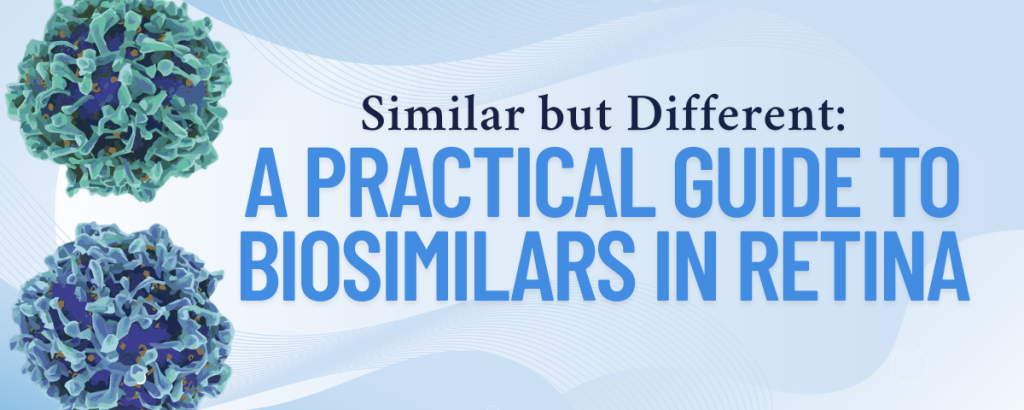
As biosimilars begin to enter the Canadian retina space, many ophthalmologists are asking: how do these agents compare to reference biologics—and what should we consider before making the switch?
Join renowned retina specialists Dr. Mike Klufas and Dr. Sophie Bakri for a 90-minute webinar that dives into the practical realities of biosimilar use—from clinical trial data to real-world adoption strategies.
📅 Date: May 29, 2025
🕒 Time: 7:00 PM ET
⏳ Duration: 90 minutes

Dr. Michael Klufas is a vitreoretinal surgeon at Wills Eye Hospital and Associate Professor of Ophthalmology at Thomas Jefferson University. He has authored over 90 publications and is a principal investigator in clinical trials on AMD, diabetic eye disease, biosimilars, and gene therapy. A graduate of Brown University and Weill Cornell, Dr. Klufas is a frequent speaker at national and international meetings.

Dr. Sophie J. Bakri is Chair of Ophthalmology at Mayo Clinic and holds the Whitney and Betty MacMillan Professorship. A vitreoretinal surgeon and Wharton MBA graduate, she has authored over 230 peer-reviewed papers and leads multiple clinical trials on retinal disease. She has received numerous accolades, including the AAO Senior Achievement Award and Retina Hall of Fame induction.
This webinar is ideal for retina specialists, general/comprehensive ophthalmologists, fellows, residents, and medical students seeking a deeper understanding of biosimilars and their role in retinal disease management.
In addition to these experts, the Biosimilars Planning Committee will be joining them in a panel discussion at the end of the webinar. We are excited to have Dr. Parnian Arjmand, Dr. Vlad Diaconita, Dr. Bryon McKay and Dr. Miral Mehta-Shounak.
Learning Objectives:
After this session, participants will be able to:
- Interpret and evaluate clinical trial data on biosimilars by analyzing key findings from pivotal ophthalmology studies and comparing their efficacy, safety, and real-world outcomes to reference biologics;
- Apply real-world evidence to clinical practice by examining case studies and identifying strategies to overcome challenges in biosimilar adoption across various ophthalmic settings;
- Navigate regulatory and practical considerations by understanding Health Canada’s biosimilar guidelines and examining reimbursement models and access barriers within Canada;
- Engage in expert-led discussions to gain insights on the evolving role of biosimilars and address common questions and concerns through interactive Q&A.
Missed the webinar?
The recording is available on demand!
This webinar would not be made possible without our sponsors. Please click here for our list of sponsors and our disclosure page.
Musculoskeletal Disorders in Ophthalmologists
The COS Ergonomics working group is pleased to share an online resource focused on musculoskeletal disorders in ophthalmologists. Developed by Queen’s University in collaboration with Weill Cornell Medicine, this online module highlights best ergonomic practices and injury prevention.
Why is this important? The physical demands of ophthalmology puts physicians at risk for work-related musculoskeletal disorders (MSDs) due to poor ergonomics.
After this module, participants will:
- Review the importance of ergonomics for ophthalmologists;
- Identify areas in the clinic and operating room that may predispose to musculoskeletal disorders;
- Learn proper ergonomic risk factor modification;
- Recognize signs and symptoms of musculoskeletal disorders, understanding the importance of early intervention.
Completion of this online module has been determined by Queen’s University to be eligible for 0.5 hours of Royal College Physicians and Surgeons of Canada Section 2: Self Learning credits.
Ready to learn more? Click below!