


The OR Microscope – The Danger Zone
By Yusuf Ahmed, Mostafa Bondok, Patrick Gooi, and R. Rishi Gupta
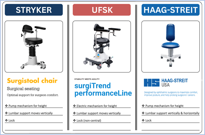
The Surgical Chair
Ergonomic positioning in the OR begins with the surgical chair. Key features to consider include adjustable height (manual or electric), vertically adjustable lumbar support with horizontal adjustability when available, and a locking mechanism that provides stability. Features of commonly used chairs are provided in the images below.
The locking mechanism, when engaged, stabilizes the surgeon and improves fine motor control by allowing relaxation of the core and large stabilizing muscles. However, this same feature can “trap” the surgeon in a suboptimal position. Once alignment is achieved and the chair locked, it is important to recognize that periodic unlocking and micro-adjustments may be necessary as the case progresses and patient positioning shifts.
Optimize and Renew Your Form
Before the case begins, a brief “form check” is advisable. Relax your body, keep your neck neutral, stack your joints, and drop your shoulders down and back. Your back should be supported by the chair’s lumbar support, and your weight evenly distributed along the seat. Position arms and legs in a comfortable, symmetric posture, with pedals at a distance that doesn’t force reaching forward or backward. Finally, aim for hips slightly higher than knees (i.e., just over 90 degrees).
Poor ergonomic habits in the OR rarely reflect neglect. More often, they arise from cognitive load, time pressure, or fatigue. Surgeons may begin a case with excellent alignment, only to find that concentration, difficult anatomy, or prolonged duration gradually erode posture. When possible, distributing complex cases across the surgical schedule may reduce cumulative musculoskeletal strain in addition to cognitive fatigue.
The OR Microscope: A Static Trap
The OR microscope introduces a unique ergonomic challenge. Once the eyes are engaged in the oculars, the body tends to become fixed in position. If alignment is non-optimal, the surgeon may remain locked in a static, non-neutral posture for extended periods. Traditional advice to “sit up straight” is incorrect. Sustaining a rigid, upright posture increases muscular effort and often leads to compensatory strain. A more effective approach is to assess where the eyes would naturally fall if the body were completely relaxed. Any sensation of effort required to maintain ocular alignment should be treated as a signal that adjustments to the microscope, chair, and/or patient positioning should be made.
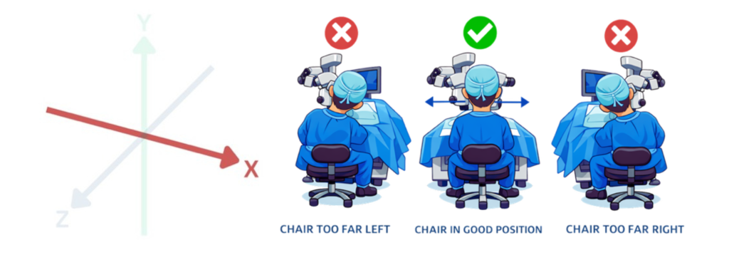
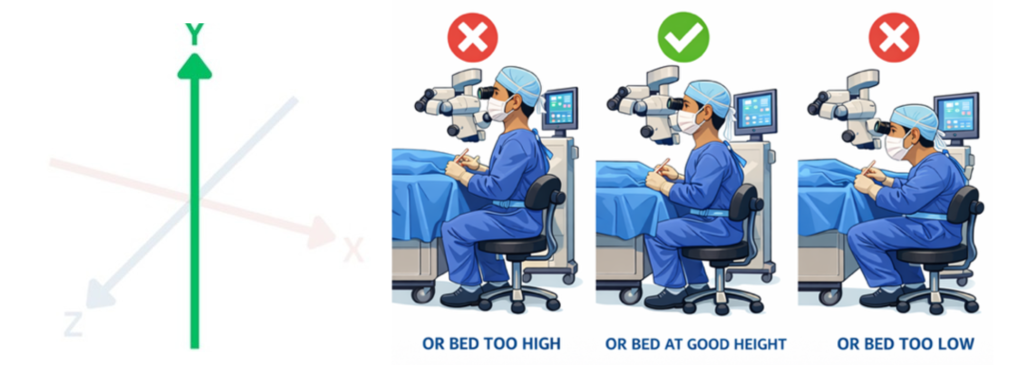
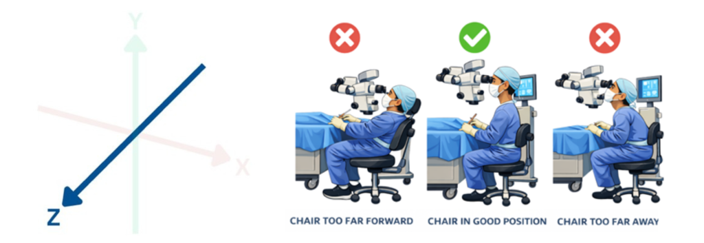
Three-Dimensional Alignment: OR microscope ergonomics can be conceptualized along three axes:
X-axis alignment – refers to left–right positioning. If the microscope is not centred relative to the surgeon, subtle lateral leaning or trunk side-bending occurs. Even small asymmetries can lead to uneven loading of paraspinal muscles and facet joints over time.
Y-axis alignment – reflects vertical positioning relative to the patient and surgical bed. When seated too low, surgeons compensate with cervical extension, increasing load on posterior cervical elements and paraspinal musculature. When seated too high, forward trunk and neck flexion predominate, increasing anterior cervical disc pressure, upper trapezius strain, and lumbar loading.
Z-axis alignment – refers to forward–backward positioning. Physical constraints imposed by chairs and pedals may prevent surgeons from positioning themselves close enough to the operative field, encouraging forward leaning. This increases lumbar disc pressure and thoracic flexion, often accompanied by compensatory cervical extension. Positioning the patient’s head closer to the edge of the headrest, when safely tolerated, can help reduce the need to reach forward.
COS Ergonomics and Injury Prevention Working Group
The Canadian Ophthalmological Society (COS) Ergonomics and Injury Prevention Working Group is committed to promoting physical wellness across all stages of ophthalmology practice. Through educational initiatives, resource development, and collaborative research, we aim to make a focus on ergonomics a standard in Canadian ophthalmic care.
Our Mission: To eliminate work-related injuries for Canadian ophthalmologists.
Our Vision: To educate every Canadian ophthalmologist on ergonomics and how best to mitigate risks.
Our Values: Respect, passion, teamwork, creativity, inclusion, and diversity.
Our Current Projects:
- Canadian Handbook for Ergonomics and Injury Prevention for Ophthalmologists
- Ergonomics Toolbox for Residents
- Resident Ergonomics Curriculum Development
- Social Media Educational Initiatives and Challenges
- Sightlines Ergonomics Educational Initiatives
- SAP: Ergonomics: Doesn’t Hurt To Think About It, But It May Hurt Not To! (Royal College MOC Section 3 Accredited)
- Webinar: We’ve Got Your Back! A COS Working Group in Ergonomics and Injury Prevention Webinar (October 2025)
- Ergonomics Podcast (Fall 2025)
- And many more…!
Ergonomics and Injury Prevention Working Group Members


Journal Articles & Editorials:
Explore the growing body of peer-reviewed research on ergonomics in ophthalmology and microsurgery.
LEARN MORE →

Magazine Articles:
Hear directly from ophthalmologists about real-world experiences, preventive tips, and system-wide challenges related to MSK injury.
LEARN MORE →

Podcasts & Videos:
Prefer to learn on the go? Explore expert-led podcasts and video content on ergonomics in ophthalmology and surgery.
LEARN MORE →

Ophthalmology Educational Series:
Engage with HIGH-YIELD accredited modules and curated training developed by national experts.
LEARN MORE →

Other Ergonomics Resources for Physicians:
Ergonomic challenges affect all healthcare providers. Learn from a broader body of literature in medicine and surgery.
LEARN MORE →
Ocular Mucous Membrane Pemphigoid Course
Location: Virtual – access the course HERE
Mucous membrane pemphigoid (MMP) is a cicatricial autoimmune disease primarily affecting mucous membranes of the conjunctiva, oral cavity, nasopharynx, oesophagus and genitals. Ocular involvement occurs in approximately 80% of cases, highlighting the importance of early diagnosis to prevent irreversible damage and potential blindness.
Authors: Melissa Lu (MD), Fady Sedarous (MD, FRCSC), Mona Harissi-Dagher (MD, FRCSC)
Learning objectives: Upon completion of this module, learners will gain a comprehensive understanding of the pathophysiology, clinical signs, diagnostic and treatment principles of ocular mucous membrane pemphigoid (OMMP), enabling them to apply this knowledge in clinical practice.
Target audience: Ophthalmology residents and ophthalmologists.
World Sight Day – ROP Resource

This year, World Sight Day (October 10, 2024) focuses on children’s eye health around the world. We want to highlight and review the existing guidelines for the screening of retinopathy of prematurity (ROP). These guidelines, developed by key organizations in ophthalmology, aim to ensure early detection and management of ROP in preterm infants to prevent life-long visual impairment and blindness. Please note that this review reflects current clinical recommendations from leading bodies and is not a COS policy or official guideline.
Screening Eligibility
Screening is recommended for infants at the highest risk of developing ROP. The criteria include:
- Gestational age: ≤30 6/7 weeks
- Birth weight: ≤1500 grams
- Neonatal risk factors: Infants born at >1500 grams or >31 weeks may still require screening (eg, infants with hypotension or those who received oxygen supplementation)
Initial screening: The first eye exam is recommended at either 31 weeks’ postmenstrual age in infants with gestational ages less than 26 6/7 weeks at birth, and at four weeks of chronological age in infants born with a gestational ages of 27 weeks or more, whichever is later.
Follow up intervals depend on the severity of the ROP observed in the initial exam.
As we recognize World Sight Day, it is important to remember that infants who develop ROP are at a higher risk of visual disorders such as: strabismus, amblyopia, high refractive errors, cataracts and glaucoma. By raising awareness and ensuring early detection and treatment, we can help protect the vision of these vulnerable children and work towards a world where everyone has access to eye care.
Reviewed by: Dr. Marie-Josée Aubin, MD (Chair), Dr. Cynthia Qian, MD (CPD Council Chair)
References:
Jefferies, A. L. (2016). Screening examination of premature infants for retinopathy of prematurity. https://cps.ca/en/documents/position/retinopathy-of-prematurity-screening
Subramanian, S., Kern, M. D., & Deegan, W. F. III. (2023). Retinopathy of prematurity guidelines. Medscape. https://emedicine.medscape.com/article/976220-guidelines
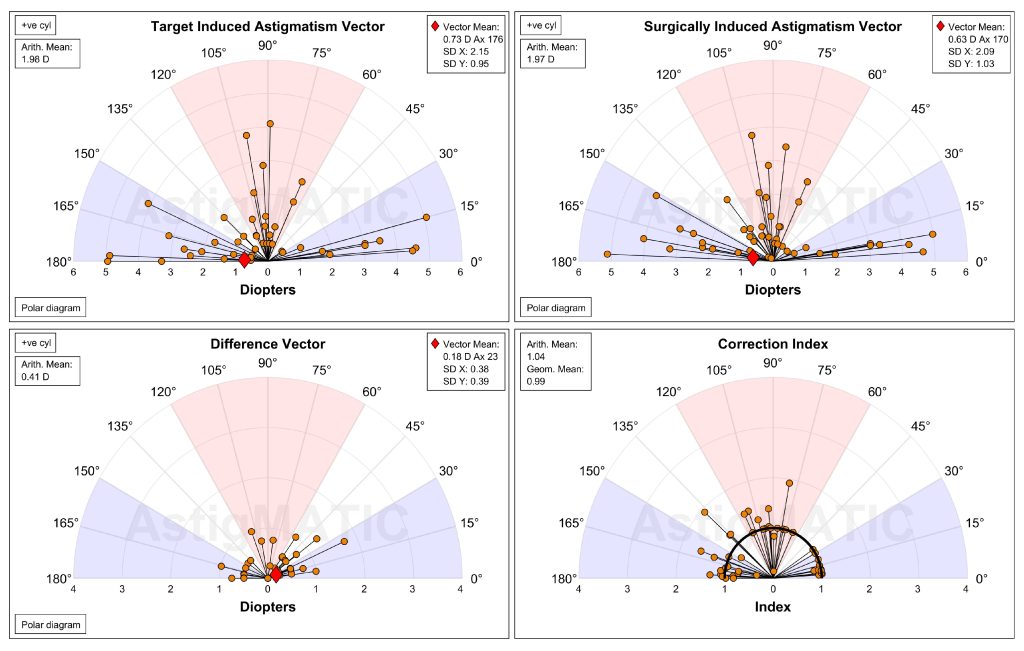
AstigMATIC: An Automated Vector Analysis Software
AstigMATIC is a free standalone application for automated standardized astigmatism vector analyses in corneal and intraocular refractive surgeries. AstigMATIC allows the simultaneous display and analysis of astigmatism magnitude and axis.
The software produces the four following graphs according to current journal standards: 1-Target-Induced Astigmatism Vector, 2-Surgically-Induced Astigmatism Vector, 3-Difference Vector and 4-Correction Index. Vector means with standard deviations are automatically calculated and displayed on the corresponding single-angle vector plots (0 to 180°). The standard graphs can be easily exported as high-resolution TIFF images for figures to use in production and presentations.
The software can be downloaded at:
https://www.lasikmd.com/media/astigmatic/index.php.
The software was thoroughly tested, peer-reviewed and published in BMC Ophthalmology, the paper about the software is available at: https://bmcophthalmol.biomedcentral.com/articles/10.1186/s12886-018-0920-1
For more information about the authors please visit their Research Unit website:
https://www.refractivesurgery.ca/
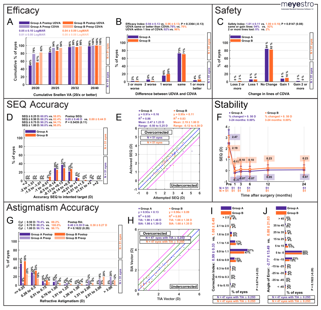
mEYEstro Automated Standard Outcomes Software
mEYEstro is a user-friendly software that automatically generates standardized graphs for refractive surgery outcomes – as prescribed by JRS, JCRS, and Cornea Journals.
The software produces the following 11 standard graphs; Efficacy: 1. Cumulative uncorrected (UDVA) and corrected visual acuity (CDVA), 2. Difference between UDVA and CDVA, Safety: 3. Change in line of CDVA, Accuracy: 4. Spherical equivalent (SEQ) to intended target, 5. Attempted vs. achieved SEQ, 6. Defocus equivalent (DEQ) accuracy, 7. Refractive astigmatism accuracy, 8. Target-induced astigmatism vs. Surgically-induced astigmatism, 9. Correction index histogram, 10. Angle of error histogram, Stability: 11. SEQ stability over time. Percent proportions, means, standard deviations, Cohen’s d effect sizes, and p-values are calculated and displayed on each graph. All graphs can be easily exported as high-resolution TIFF images for figures to use in scientific manuscripts and presentations.
The software can be downloaded at https://www.lasikmd.com/media/meyestro/index.php, and a tutorial is available at https://www.youtube.com/watch?v=NFlRRHx6ZaI.
Please note that our software was recently thoroughly tested, peer-reviewed and published in BMC Ophthalmology, the paper about the software is available at: https://bmcophthalmol.biomedcentral.com/articles/10.1186/s12886-023-02904-6
Organ Culture of Corneas and its Role in Eye Banking Economics
Date: Wednesday, November 30, 2022
Time: 4:00-5:30pm ET.
Presented by Dr. Graeme Pollock, Director of Lions Eye Donation Service, Centre for Eye Research Australia
Hosted by Gary Rockl, Tissue Innovation Specialist from Héma-Québec
The presentation will examine the technical aspects of organ culture of donor corneas and the benefits that may accrue through its adoption. It places this within the context of the forces affecting supply, demand and distribution of corneas in Australia.
• What are the technical requirements of organ culture?
• What are the benefits and disadvantages of the system?
• How does Australia approach national networking, self-sufficiency and sustainability in the provision of corneas for transplantation?
About the Featured Presenter:

Dr. Graeme A. Pollock, BSc (Hons) GCUT MPH PhD OAM
Dr Pollock is the Director of Lions Eye Donation Service at the Centre for Eye Research Australia, Melbourne. He has spent the past 30 years involved in eye banking at local, national, and international levels. Having been trained in pathology at the University of Melbourne, His association with donation and transplantation extends back to the 1980’s while working in organ preservation for transplantation. He completed his doctorate in this field at the University of Queensland and University of Cambridge. Graeme also holds a master’s degree in public health majoring in Health Administration from Monash University, and a Post-Graduate Certificate in University Teaching from the University of Melbourne.
His interests include medical standards and guidelines in the field of eye banking. He is a past Chair of the Eye Bank Association of Australia and New Zealand, past-President of the Australasian Transplant Coordinators Association, a member of the Medical Advisory Board of the Eye Bank Association of America and a founding member of the Global Alliance of Eye Bank Associations. In 2019 he was awarded an Order of Australia for service to corneal transplantation and education.
This meeting will be recorded for those who are unable to attend.

ESCRS Launches Online IOL Calculator
The developers’ goal was to create a free online tool that helps ophthalmologists aggregate the major online IOL calculators in one site, allowing them to get multiple results with only one data entry session.
“This will reduce typing mistakes, provide a better view, and allow users to compare results, eventually helping to get better refractive outcomes,” Dante Luis Buonsanti told EuroTimes.
The developers used a technique called web scraping. This is the sort of thing seen when using a hotel or air travel search engine. Web scraping uses bots to extract content from several websites and sends it to a single site. The user can then compare results from seven current calculators: Barrett Universal II, Cooke K6, Evo, Hill-RBF, Hoffer QST, Kane, and PEARL GDS. The site also “scraped” a comprehensive list of IOLs with optimized constants from IOL.com.
The ESCRS online IOL calculator is now live at iolcalculator.escrs.org.
Free slit lamp breath shields for your practice
March 25, 2020
Zeiss is giving all ophthalmologists 2 free slit lamp shields for free.
Access Details
To receive your free breath shield fill out the information required on:
https://www.zeiss.com/meditec/int/c/slit-lamp-breath-shields.html?vaURL=www.zeiss.com/breathshield