


Explore the Practice Resource Centre
EXPLORE THE PRACTICE RESOURCE CENTRE – we are pleased to share the new PRC promotional video. In just over two minutes, we highlight the benefits of the site for seasoned practitioners and those just starting their career, how to easily submit ideas and resources, how to create a custom reading list, and much more.
Glaucoma OCT Interpretation: 101
Glaucoma OCT Interpretation is an online, Section 3 accredited course available at EyeCarePD.com. A game-based approach is applied to learning OCT interpretation. This course covers commonly encountered OCT presentations of glaucoma as seen on standard optic nerve head and retinal nerve fiber layer scan protocols with the purpose of improving interpretation skills using perceptual learning strategies.
Getting started is easy:
- Using the Google Chrome browser, navigate to https://eyecarepd.com/catalog/glaucoma-group/
- Select Glaucoma OCT Interpretation 101 (COS Accredited) and add the product to the cart
- At checkout, use the coupon code ECPDCOSGLAUCOMA to set the price to 0.
- Your course is now available under My Courses at the top of the screen.
Learning Objectives:
- Identify segmentation errors and their role in OCT scan interpretation
- Utilize varying sections of the OCT report including tabular data, sector analysis and graphical displays
- Interact with expert interpretation in order to compare their findings in selected cases
Accreditation
This activity is an Accredited Self-Assessment Program (Section 3) as defined by the Maintenance of Certification program of the Royal College of Physicians and Surgeons of Canada. This web-based lesson was approved by the COS on October 23rd, 2020 and expires October 2023. Remember to visit MAINPORT to record your learning and outcomes. You may claim a maximum of 1 hour (credits are automatically calculated).
This activity was co-developed with EyeCarePD and the Canadian Ophthalmological Society (COS) and was planned to achieve scientific integrity, objectivity and balance.
Access Details
To participate in this course visit https://eyecarepd.com/catalog/glaucoma-group/
2026 COS Hot Topics ePoster Presentation Webinar: Recording Now Available
Did you miss the COS Hot Topics Webinar? The recording is now available!
Held on June 24, 2026, the webinar featured a dynamic lineup of top-ranked ePoster presentations from the 2026 COS Annual Meeting & Exhibition, showcasing innovative research and emerging trends in ophthalmology. The session highlighted exciting developments in imaging, retinal disease, cornea, artificial intelligence, and therapeutic advancements.
Thank you to Dr. Adam Muzychuk for moderating an engaging and insightful discussion, and to all presenters for sharing their outstanding work.
Featured presentations included:
- Marwa Al Ghafri
Short-Term Outcomes of Switching to Faricimab in nAMD and DME Patients with Suboptimal Responses to Aflibercept - Aya Benabdelhak
Tracking Corneal Nerve Regrowth After Neurotization: A Validated, Standardized IVCM Methodology - Jiwon Hwang
Longitudinal Assessment of Growth and Melanin Change in Choroidal Nevi with Polarization Diversity Optical Coherence Tomography (PD-OCT) - Tina Felfeli
Intraocular Fluid Cytokine Profiles as Biomarkers for Retinal Disease Prediction: A Machine Learning Approach - Devina Ramesh
Patient Preference for Topical Antisepsis for Intravitreal Injection: A Prospective Clinical Study - Zhao Feng
Study of Ophthalmic Radiation Therapy Toxicity: Classification System for Radiation Retinopathy - Godfrey Wong
External Validation of the Multifocal Electroretinogram Classification Interface (MERCI), a Machine Learning Algorithm for Hydroxychloroquine Toxicity - Tasha Miller
Redistribution of Corneal Endothelial Cells Following Descemet’s Stripping Only for Fuchs Endothelial Corneal Dystrophy - Lia Huo
A Gene-Agnostic Approach: Sustained Delivery of Rod-Derived Cone Viability Factor Protects Human and Rodent Cones in Retinitis Pigmentosa - Sohel Somani
SPECTRUM: Data From a Global Real-World Study of Aflibercept 8mg in Patients with Treatment-Naïve or Previously Treated Diabetic Macular Edema
Watch the recording to explore cutting-edge research shaping the future of ophthalmology.
2026 COS Film Festival Recordings Now Available
Did you miss the inaugural COS Film Festival at the 2026 COS Annual Meeting and Exhibition in Montréal? Or want to revisit some of the outstanding submissions?
We are pleased to share recordings of all 13 videos presented during the first-ever COS Film Festival, held during the 2026 Annual Meeting and Exhibition. This new event was created to celebrate innovation, foster learning, and showcase the exceptional talent within the Canadian ophthalmology community.
The Film Festival featured a wide range of high-quality surgical and educational videos highlighting innovative techniques, complex cases, and valuable clinical insights across ophthalmology. Presentations were evaluated based on quality, importance, and educational value, with awards recognizing outstanding submissions.
COS Film Festival Video Lineup
Videos are listed in final ranking order.
Geoffrion, Dominique
Rudnisky, Chris
Gupta, R. Rishi
Strube, Yi Ning
Wandzura, Adam
Wandzura, Adam
Sverdlichenko, Irina
Abdelnour, Patrik
Jakubowska, Weronika
You, Jia Yue
Punja, Karim
Florido, Antonio
Schell, Carson
Advanced Therapeutic Options for Ocular Surface Disease: Blood-Derived Products, Insulin Eye Drops, and Human Amniotic Membrane
COS Practice Resource Centre General Information for Patients and Providers
Ergonomics and Injury Prevention Webinar
Save the Date!
Date: Thursday, October 8, 2026, 7:00 pm ET
Title: Keeping You in the Loupe: A Closer Look at Ergonomics in the Operating Room
Intravenous Acetazolamide Shortage
The Atlantic Eye Symposium
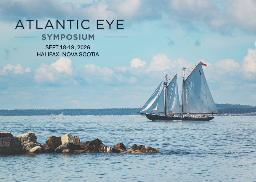
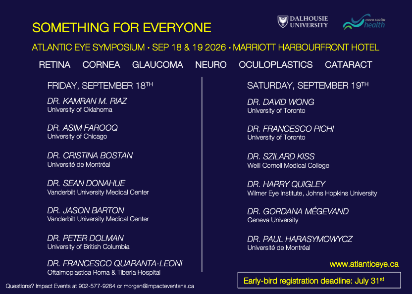
The Atlantic Eye Symposium is an accredited educational conference geared to address the needs of the comprehensive ophthalmologist. There will be 13 visiting speakers covering a variety of topics including cataract, cornea, retina, glaucoma, neuro-ophthalmology, and oculoplastics.
This two-day meeting will be held on the Halifax waterfront at the Harbourfront Marriott Hotel on September 18th and 19th, 2026. The informal setting will facilitate dialogue and interaction with the speakers and your colleagues. You can expect innovative talks and exhibitions addressing current issues in the field of ophthalmology.
EyeNovation Webinar: INTRA-KER: A Novel Concept for a Hybrid Keratoprosthesis
Did you miss the webinar? Not to worry, a recording is available for you below!
Our EyeNovation webinar featured Dr. Massimo Busin, who presented on INTRA-KER: A Novel Concept for a Hybrid Keratoprosthesis.
This session explored the concept of INTRA-KER, a hybrid keratoprosthesis that integrates a synthetic optical device with a biologic scaffold, offering practical insights and clinical perspectives relevant to ophthalmologists and trainees.
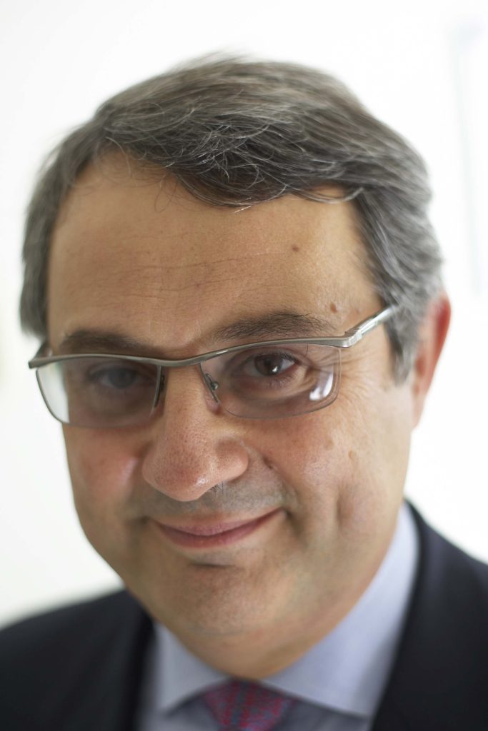
Massimo Busin, MD, is the chairman of the Department of Ophthalmology at “Ospedali Privati Forlì” in Forlì (Italy) and is also a Professor of Ophthalmology at the University of Bonn (Germany), as well as and Adjunct Associate Clinical Professor at LSU (New Orleans, USA). Since 2017 he has been appointed Professor of Ophthalmology and Director of the Residency program of the Department of Ophthalmology in Ferrara (Italy)
Dr. Busin has received from the AAO (American Academy of Ophthalmology) the “Honor Award” in 1993, the “Senior Honor Achievemt” award in 2003, the “Life Achievement” award in 2013 and the “Special Recognition Award” in 2015; from SOI (Società Oftalmologica Italiana) the Gold Medal “Maestro dell’Oftalmologia” in 2012. He has delivered several eponimal lectures and has also received several prizes for both videos and lectures delivered at international meetings, among which the AAO, the ESCRS, the DOC, the ASCRS, the SOI. He has authored more than 250 peer-reviewed papers, and 24 book chapters.
Learning Objectives
By the end of this session, participants will be able to:
- Understand the foundational concept, key surgical principles as well as clinical indications of INTRA-KER
CanMEDS Role: Medical Expert
Representation Circle: Surgical Learning, Research and Mentorship – Recording
Did you miss the webinar? Not to worry, a recording is available for you below!
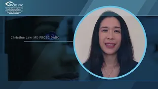
Our inaugural Representation Circle webinar featured Dr. Prithvi Mruthyunjaya, who presented on Surgical Learning, Research and Mentorship.
This interactive session explored mentorship, surgical learning, and research development in ophthalmology training, while creating space for meaningful discussion and shared experiences across the ophthalmology community. The session also included a live Q&A with participants.
Dr. Prithvi Mruthyunjaya is the Alan Adler Professor of Ophthalmology, member of the Vitreoretinal Surgery Service, and Director of Ocular Oncology at the Byers Eye Institute at Stanford. As Director of the Vitreoretinal Surgery and Medical Retina Fellowships, he has helped build a highly regarded academic-focused retina training program and has trained more than 50 retina and ocular oncology fellows over the past 20 years. He has coauthored over 230 peer-reviewed publications and is internationally recognized for his contributions to surgical education, mentorship, and vitreoretinal training.
Dr. Awwad serves as Associate Editor of the Journal of Refractive Surgery and sits on the Editorial Board of the American Journal of Ophthalmology. His work has earned numerous international distinctions, including the George Waring Medal for Editorial Excellence and the AAO Achievement Award. He has also contributed innovative analytical tools and formulas to major ophthalmic platforms and holds patents in artificial intelligence and computer vision related to ophthalmic surgery.
Learning Objectives
By the end of this session, participants were able to:
- Review elements of organizing surgical learning in residency
- Understand concepts of research development in residency
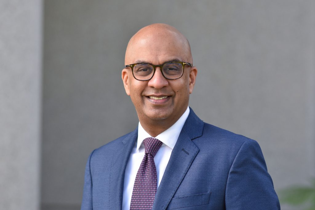
EyeNovation Webinar: The Evolving Landscape of CAIRS – Recording
Did you miss the webinar? Not to worry, a recording is available for you below!
Our EyeNovation webinar featured Dr. Awwad, who presented on The evolving landscape of CAIRS.
This session explored the latest developments in CAIRS, including evolving surgical techniques, preparation methods, and clinical outcomes, offering valuable, practice-oriented insights for ophthalmologists and trainees.
Dr. Shady Awwad is Professor of Ophthalmology and Head of the Cornea & Refractive Surgery Division at the American University of Beirut Medical Center, where he also founded the Refractive Surgery Fellowship Program. He is internationally recognized for his expertise in refractive and keratoconus surgery and has authored more than 130 peer-reviewed publications.
Dr. Awwad serves as Associate Editor of the Journal of Refractive Surgery and sits on the Editorial Board of the American Journal of Ophthalmology. His work has earned numerous international distinctions, including the George Waring Medal for Editorial Excellence and the AAO Achievement Award. He has also contributed innovative analytical tools and formulas to major ophthalmic platforms and holds patents in artificial intelligence and computer vision related to ophthalmic surgery.
Learning Objectives
By the end of this session, participants will be able to:
- Introduction to CAIRS, their outcomes and their mechanism of action
- Insights into the changing trends in CAIRS preparation, insertion, and surgical techniques.